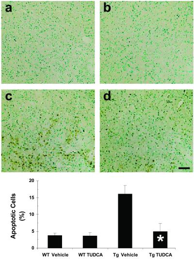
Fig 1.
TUDCA reduces striatal apoptosis in HD Tg mice. Representative striatal photomicrographs of TUNEL-stained sections are shown for each treatment group. Quantitation of apoptotic cells is indicated in the bar graph. Control Tg HD mice (c) exhibited significantly increased proportions of apoptotic vs. total cells compared with vehicle (a) and TUDCA-treated (b) wt control mice. TUDCA-treated R6/2 mouse striata (d) contained significantly fewer apoptotic cells compared with untreated Tg mice. Sections are counterstained with methyl green. *, P < 0.01 for Tg TUDCA vs. vehicle. (Scale bar, 100 μm.)

