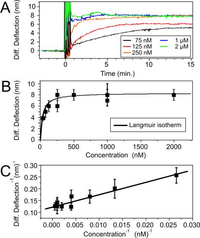
Figure 3.
(A) The target concentration-dependent studies show the time scale for the cantilever to equilibrate to different concentrations of oligonucleotides. Systematic hybridization measurements were performed on a single cantilever, and sequence-specific differential signals were extracted by using a reference cantilever. The spikes in the differential signal result from the injection of the solutions into the fluid chamber, but rapid equilibration is reached within a few minutes. Depending on injection speed and volume, the dehybridization of the double helix was achieved by a urea injection. (B) Concentration-versus-deflection plot of many levers shows that the hybridization of the oligonucleotides follows the Langmuir isotherm model (15). (C) Langmuir plot (15) of the data in B.

