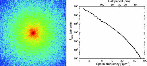
Fig. 1.
Soft x-ray diffraction pattern of a freeze-dried yeast cell. (Left) The assembled pattern shown represents the summation of several diffraction patterns, with a total exposure of 65 seconds to 750-eV x-rays. The assembled pattern covers 1200 × 1200 pixels of the 1340 × 1300 original array and extends to a spatial frequency, or diffraction angle divided by wavelength, of 48 inverse micrometers at the edges and 68 inverse micrometers at the corners. A spatial frequency of 48 inverse micrometers at the edges corresponds to a half-period pixel size in real space of 10 nm. The speckles in the diffraction pattern have a consistent size associated with the inverse of the size of the yeast cell. (Right) Power spectrum as a function of spatial frequency.

