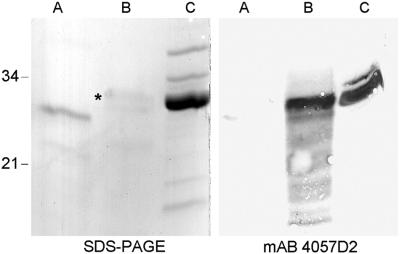
Fig 5.
SDS/PAGE and immunoblot analyses of rMS-LBP, chemically methylated rMS-LBP, and nMS-LBP. rMS-LBP (A), rMS-LBP subjected for 15 min to chemical lysine methylation (B) and nMS-LBP (C) were analyzed by SDS/PAGE followed by Coomassie blue staining (Left) or by immunoblotting by using mAb 4057D2 (Right). The sizes of the Mr markers given in kDa are shown in the left margin, and the band corresponding to chemically methylated rMS-LBP is indicated by *.

