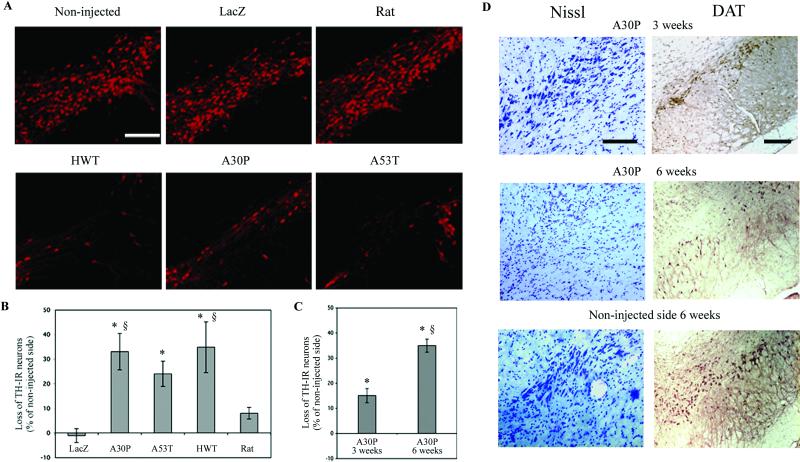
Fig 2.
Dopaminergic-specific marker (TH, DAT) expression in the substantia nigra of rats injected with lentiviral vectors encoding for wild-type (HWT) or mutated (A30P, A53T) human and wild-type rat α-syn. (A) Overexpression of normal or mutant human α-syn significantly decreases the number of TH-IR neurons. No significant loss of TH expression was observed with the lentivirus encoding for rat α-syn or the reporter protein β-galactosidase (LacZ). (B) Histograms representing the loss of TH-IR nigral neurons at 5 mo relative to the contralateral side in rats unilaterally injected with the different lentiviral constructs. (C) The loss of dopaminergic neurons was quantified at 3 and 6 weeks for the A30P mutant and compared to Lenti-LacZ- or Lenti-rat α-syn-injected animals at 5 mo. The A30P lentiviral-mediated lesion represents a time-dependant process occurring within 6 weeks. (D) Nissl and DAT staining for A30P expressing animals at 3 and 6 weeks confirming the loss of dopaminergic cells over time. Values refer to means ± SEM; n = five animals per group; *, P < 0.005; §, P < 0.05; *, compared to lenti-lacZ-injected animals; §, compared to lenti-rat α-syn-injected animals at 5 mo. (Scale bars = 200 μm.)

