Fig 4.
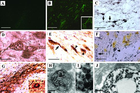
Neuropathology induced by lentiviral-mediated expression of α-syn. (A) No fluoroJade B labeled neurons were detected in both Lenti-LacZ-injected animals and noninjected side of animals expressing human α-syn. (B) Scattered FluoroJade B-positive neurons detected in the substantia nigra of rats expressing A30P α-syn. Higher magnification shows a degenerating neuron with FluoroJade B labeling in their perikaria and neurites. (C and D) α-Syn staining revealed that transduced neurons develop dystrophic neurites (C, arrows) and abnormal α-syn immunopositive swollen structures (D) with axonal and cell body distribution. (E) Nigral neurons abnormally accumulate cytoplasmic α-syn-immunopositive aggregates in cell bodies and neurites in rats overexpressing A30P α-syn. To confirm the presence of inclusions rather than a particular cellular sublocalization of α-syn, a silver staining (dark deposits) was performed alone (F) or in combination with α-syn staining (red-brown color) (G). Cytoplasmic inclusions (arrowheads) and extensive neuritic pathology (arrows) are detected with both methods. Higher magnification of a double-stained aggregate reveals that the inclusions contain abundant α-syn. (H–J) Ultrastructural analysis of A30P α-syn expressing nigral neurons. Abundant α-syn immunoreactive aggregation was detected in both the axon (H and I) and cell body (J) of nigral neurons expressing human α-syn. These cytoplasmic structures are mainly present as scattered granular microaggregates, which are occasionally associated with mitochondria or synaptic vesicles (H) and nuclear or cytoplasmic membrane (J). A degenerating cell with clustered aggregates in its perikaria reveals an important loss of organelles in its cytoplasm and a disorganized nuclear membrane (J). The size of the cell body and the presence of a varicosity full of synaptic vesicules observed (on the top) in close apposition with α-syn-positive structures support that this degenerating cell is a neuron. [Scale bars = 50 μm (A, B, and G); 25 μm (C, E, and F); 8 μm (D); and 0.3 μm (H and J).]

