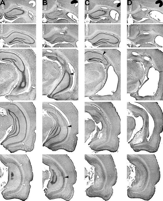
Fig 1.
Representative cresyl violet stains of intact neuronal cell bodies at five coronal levels through the hippocampus after sham surgery (A) or ibotenate lesions of the dorsal hippocampus (B), ventral hippocampus (C), or entire hippocampus (D). Filled arrowheads, borders between lesioned and healthy tissue; open arrowheads, borders of the subiculum.

