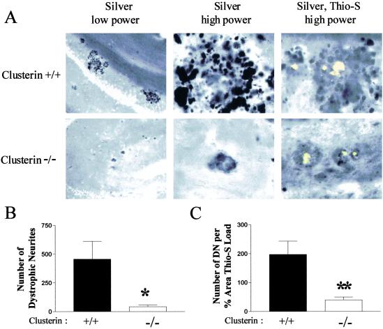
Fig 3.
Dissociation between amyloid plaques and neurite toxicity in PDAPP+/+, clusterin−/− mice. (A) Brain sections from 12-month-old PDAPP+/+, clusterin+/+ and PDAPP+/+, clusterin−/− mice were labeled with the de Olmos silver stain with or without thioflavine-S (Thio-S) to identify the neuritic dystrophy associated with the fibrillar amyloid. Vast numbers of dystrophic neurites (DN) were observed in the locale of thioflavine-S-positive deposits in PDAPP+/+, clusterin+/+ mice (Upper) at low and high power. Little neuritic dystrophy surrounded thioflavine-S-positive deposits in the PDAPP+/+, clusterin−/− mice (Lower). (B) PDAPP+/+, clusterin−/− mice had significantly fewer dystrophic neurites (mean ± SEM: 42.9 ± 13.8, n = 15) in three equally spaced sections than PDAPP+/+, clusterin+/+ mice (456.6 ± 155.2, n = 13). *, P = 0.0083. (C) The number of dystrophic neurites normalized to the percent area of the hippocampus covered by thioflavine-S was significantly decreased (5-fold) in PDAPP+/+, clusterin−/− mice (mean ± SEM: 40.0 ± 10.1, n = 15) compared with the PDAPP+/+, clusterin+/+ mice (197.3 ± 45.8, n = 13). **, P = 0.0014.

