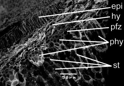
Fig 2.
Scanning electron microscopy (SEM) micrograph of a cross section through a lignified rind from C. moschata showing the hypodermis (hy), stone cells (st), phytoliths (phy), and phytolith forming zone (pfz). epi, epicarp (outer surface) of the rind. The stone cells are elongated, leading to elliptical phytoliths with elongated meoscarp-derived hemispheres and impressions (phytolith on the right) unlike those in Fig. 1.

