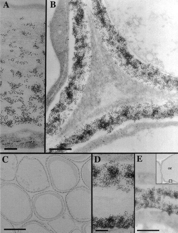
Figure 3.
Immunolocalization of β-glucans in mesophyll cells and epidermis of developing maize coleoptiles using 10 nm colloidal gold-conjugated secondary antibody. A, The gradient of β-glucans immunolocalized in the outer wall of the epidermis. The cuticle is at the top of the figure, cytoplasm at the base. Scale bar represents 200 nm. B, β-Glucan epitopes are excluded from the cell corner formed by three mesophyll cells. Scale bar represents 500 nm. C, Low magnification image showing the immunostain in a sharply defined region of the wall closest to the plasma membrane of cells. Scale bar represents 5 μm. D, β-Glucans immunolocalized in two neighboring mesophyll cell walls but excluded from the middle lamella and older regions of wall. Scale bar represents 200 nm. E, β-Glucans immunolocalized uniformly across the inward-facing wall of the epidermis. Scale bar represents 200 nm.

