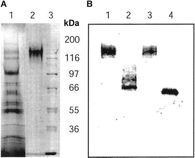
Figure 2.
SDS-PAGE and western analysis of the purified recombinant Δ1-90Cel16. Proteins were separated on a 10% (w/v) polyacrylamide gel by SDS-PAGE and stained with Coomassie Brilliant Blue (A) or transferred onto a polyvinylidene difluoride membrane and probed with a Cel16 antiserum (B). Lane A1, Crude extract from P. pastoris culture broth after ultrafiltration; lane A2, purified Δ1-90Cel16; lane A3, molecular mass protein marker; lane B1, purified Δ1-90Cel16 incubated with buffer; lane B2, purified Δ1-90Cel16 incubated with peptide-N-glycosidase-F (PNGaseF); lane B3, purified Δ1-90Cel16 incubated with buffer; and lane B4, purified Δ1-90Cel16 incubated with endoglycosidase-F (EndoF)/PNGaseF.

