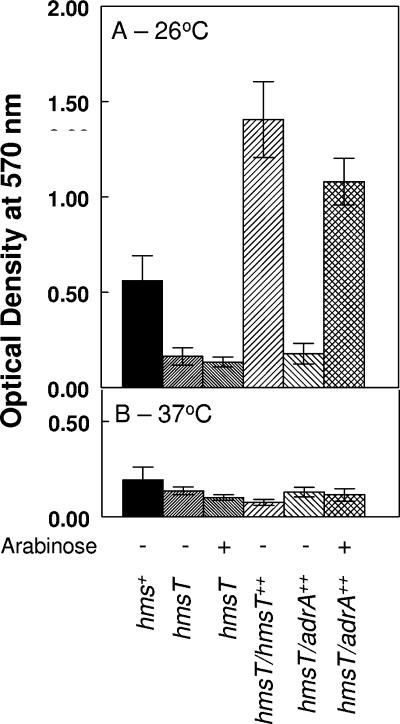
FIG. 1.
Crystal violet staining of Y. pestis cells attached to borosilicate glass test tubes after growth at 26°C (A) or 37°C (B) in media containing either glucose or arabinose. After staining with crystal violet was performed, the dye was solubilized with ethanol-acetone and optical densities were measured at 570 nm. KIM6+, hms+; KIM6-2051(pBAD30)+, hmsT; KIM6-2051(pAHMS14)+, hmsT/hmsT++; KIM6-2051(pWJB30)+, hmsT/adrA++. Values are averages from two or more independent experiments, with error bars indicating standard deviations.

