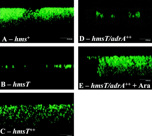
FIG. 2.
Confocal laser scanning microscopy images of green fluorescent protein-expressing Y. pestis KIM6+ (hms+), KIM6-2051+ (hmsT), KIM6-2051(pAHMS14)+ (hmsT++), and KIM6-2051(pWJB30)+ (hmsT/adrA++) cells attached to a glass coverslip after overnight growth at 26°C in the absence (A to D) or presence (E) of arabinose. Representative areas from one of three independent replicate studies are shown in the xzy plane.

