FIG. 1.
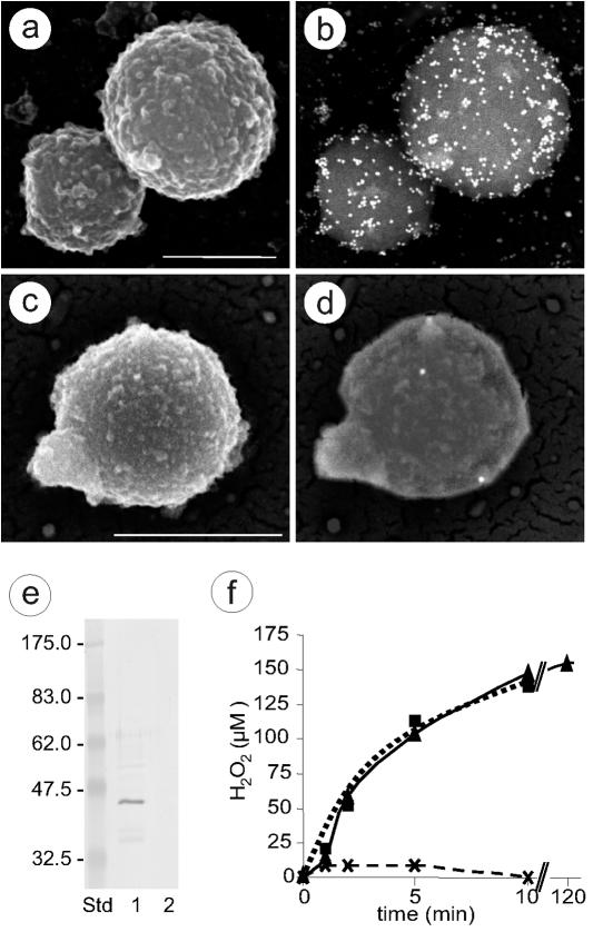
Location and activity of l-α-glycerophosphate oxidase. (a to d) SEM photographs showing immunogold labeling of M. mycoides subsp. mycoides SC strain Afadé incubated with IgG from anti-GlpO (a and b) and preimmune serum (c and d). Secondary electron micrographs show the cell surface (a and c), and back-scattered electron micrographs reveal 15-nm colloidal gold-conjugated secondary antibody (b and d). Scale bar, 500 nm. (e) Immunoblot analysis of Triton X-114-fractionated total antigens of M. mycoides subsp. mycoides SC strain Afadé with GlpO antiserum. Lane 1, detergent phase, 20 μg of proteins; lane 2, aqueous phase, 20 μg of proteins; Std, molecular mass standard. (f) Hydrogen peroxide production of M. mycoides subsp. mycoides SC strain Afadé after the addition of 100 μM glycerol. The data shown are the mean values of five independent measurements. Standard deviations of the individual measurements were below 5% of the mean values. Triangles and solid line, untreated M. mycoides subsp. mycoides SC; crosses and dashed line, M. mycoides subsp. mycoides SC pretreated with Fab fragments from anti-GlpO IgG; squares and dotted line, M. mycoides subsp. mycoides SC pretreated with Fab fragments from anti-LppC.

