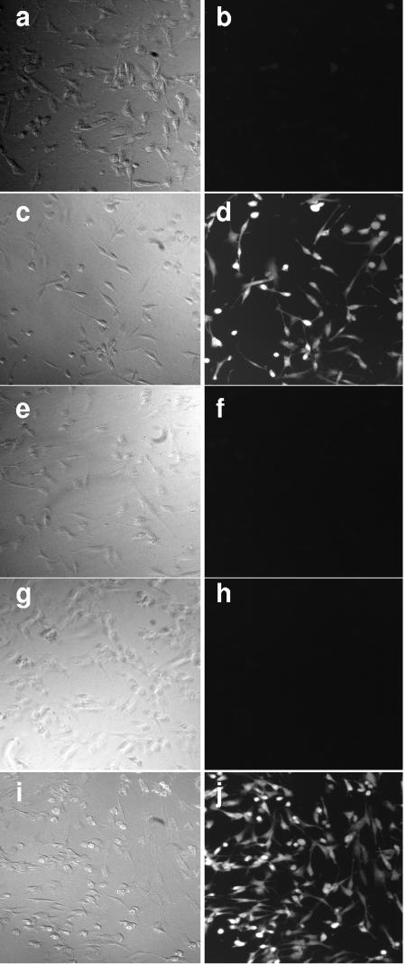
FIG. 3.
Detection of intracellular H2O2 and ROS in ECaNEp cells infected with M. mycoides subsp. mycoides SC strain Afadé at an MOI of 500 mycoplasmas per cell or treated with H2O2. Phase-contrast micrographs (a, c, e, g, and i) and fluorescence micrographs (b, d, f, h, and j) of ECaNEp cells as follows: 20 min after infection with M. mycoides subsp. mycoides SC strain Afadé in the absence of glycerol (a and b); infected with strain Afadé in medium supplemented with glycerol (c and d); infected in medium supplemented with glycerol with strain Afadé pretreated with Fab fragments from anti-GlpO IgG (e and f); pretreated with N-acetyl-l-cysteine and then infected with strain Afadé in medium with glycerol (g and h); incubated for 20 min in medium supplemented with 4.4 mM H2O2 (i and j).

