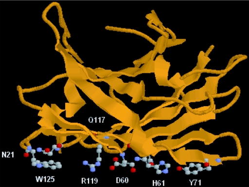
FIG. 2.
Model structure of CbpA-CBD. The structure was modeled with the program Swiss-Model version 3.5 at the Expasy server (11). The structure of CipA-CBD from C. thermocellum (18) was used as the modeling template. The structure model was visualized with the cartoon mode of the Protein Explorer software (8). The similarity of amino acid sequences between the CbpA-CBD and the CipA-CBD was 49.7%. The amino acid residues in the planar strip and the anchoring residues are visualized with the ball-and-stick mode.

