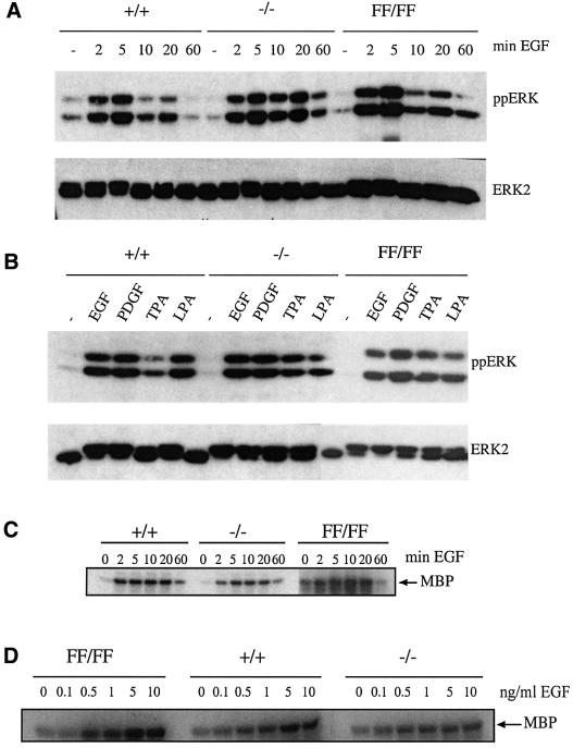
Fig. 5. ERK activation. (A) Stimulation of ERK phosphorylation in raf-1+/+, raf-1–/– and raf-1FF/FF MEFs over a time course of EGF treatment. (B) Stimulation of ERK phosphorylation in raf-1+/+, raf-1–/– and raf-1FF/FF MEFs following treatment with different stimuli for 10 min. The blots in (A) and (B) were incubated with an anti-phosphoERK antibody (top panels) and an anti-ERK2 antibody (bottom panels) to control for protein loading. (C) ERK activation in raf-1+/+, raf-1–/– and raf-1FF/FF MEFs over a time course of EGF treatment as measured by the immunocomplex MBP kinase assay. (D) ERK activation in raf-1+/+, raf-1–/– and raf-1FF/FF MEFs following stimulation with different concentrations of EGF for 10 min as measured by the MBP kinase assay.

An official website of the United States government
Here's how you know
Official websites use .gov
A
.gov website belongs to an official
government organization in the United States.
Secure .gov websites use HTTPS
A lock (
) or https:// means you've safely
connected to the .gov website. Share sensitive
information only on official, secure websites.
