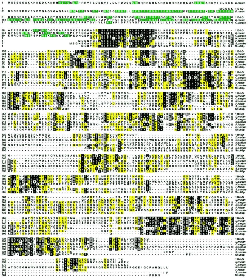
Fig. 2. Sequence alignment of the human 110 kDa protein with homologous proteins. The human 110 kDa protein is shown aligned by the Clustal method with the corresponding proteins from D.melanogaster, C.elegans, N.crassa, P.falciparum, S.pombe and S.cerevisae (Snu66p). Residues identical in at least four sequences are shown on a black background, and conserved residues, grouped as in Figure 1, are shaded yellow. RS, RD and RE dipeptides in the N-terminal part of the sequence are in green.

An official website of the United States government
Here's how you know
Official websites use .gov
A
.gov website belongs to an official
government organization in the United States.
Secure .gov websites use HTTPS
A lock (
) or https:// means you've safely
connected to the .gov website. Share sensitive
information only on official, secure websites.
