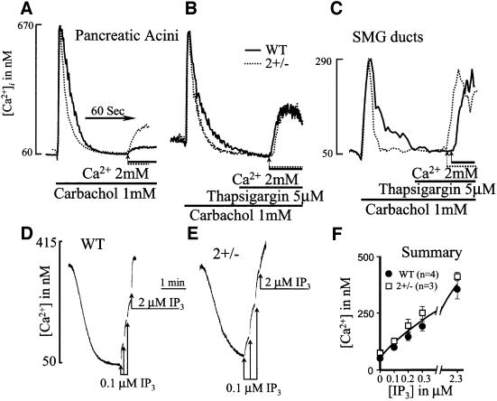
Fig. 2. Measurement of Ca2+ release and Ca2+ influx in cells from wild-type and SERCA2+/– mice. In (A–C), cells from wild-type (solid lines) or SERCA2+/– mice (dashed lines) were incubated in Ca2+-free medium prior to stimulation with 1 mM carbachol. After reduction of [Ca2+]i to a stable level, the cells were incubated with a solution containing 2 mM CaCl2 to estimate the rate of Ca2+ influx. In (B) and (C), the cells were treated with 5 µM thapsigargin to inhibit any remaining SERCA pump activity prior to initiation of Ca2+ influx. To measure directly Ca2+ uptake and release from internal stores, pancreatic acini from wild-type (D and F) or SERCA2+/– mice (E and F) were permeabilized by addition of cells to an SLO-containing medium. When medium Ca2+ was reduced to a stable level, incremental concentrations of IP3 were added to estimate the extent and potency of IP3-mediated Ca2+ release.

An official website of the United States government
Here's how you know
Official websites use .gov
A
.gov website belongs to an official
government organization in the United States.
Secure .gov websites use HTTPS
A lock (
) or https:// means you've safely
connected to the .gov website. Share sensitive
information only on official, secure websites.
