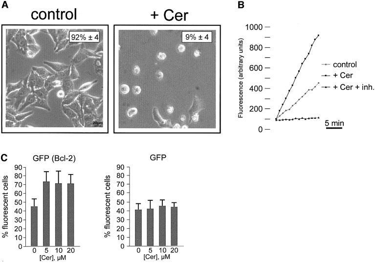
Fig. 1. C2 ceramide induces apoptotic cell death. (A) HeLa cells were maintained in KRB supplemented with 1 mM Ca2+ (1 mM Ca2+/KRB), and challenged with 10 µM ceramide (+ Cer). The number of viable cells was determined by phase contrast microscopy after 16 h. The percentage of living cells is reported in the upper right corner. Ceramide induces caspase activation in HeLa cells (B). Cells were incubated with ceramide for 16 h, and caspase-3-like activity of cell lysates was measured as detailed in Materials and methods, and expressed as fluorescence arbitrary units. The caspase inhibitor Ac-DEVD-CHO (inh.) has also been used to show specificity of the caspase-3-like activity. Bcl-2 overexpression protects cells from C2 ceramide-induced cell death (C). HeLa cells were cotransfected with Bcl-2 and with mtGFP expression plasmids. Thirty-six hours after transfection, cells were challenged with increasing ceramide concentrations (from 0 to 20 µM) and viability was assessed by microscope count of living GFP-expressing cells (see text for details). mtGFP alone does not affect cell viability. In order to eliminate possible errors due to the detachment of dead cells during the transfer of the coverslip to the chamber of the fluorescent microscope, the effect of Bcl-2 expression on cell survival was evaluated, and expressed as the percentage of fluorescent cells in the microscope field. Data are averages ± SD of triplicate determinations from experiments repeated at least five times.

An official website of the United States government
Here's how you know
Official websites use .gov
A
.gov website belongs to an official
government organization in the United States.
Secure .gov websites use HTTPS
A lock (
) or https:// means you've safely
connected to the .gov website. Share sensitive
information only on official, secure websites.
