
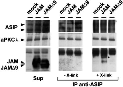
Fig. 5. ASIP and JAM associate in mammalian epithelial cells. MDCK II cells transfected with empty vector, expression vector encoding Flag-JAM or Flag-JAMΔ9 were subjected to a Ca2+ switch for 1 h, then lysed in the presence or absence of a chemical cross-linker. Lysates were incubated with affinity-purified ASIP antibodies, and the resulting immunocomplexes were analysed using the antibodies indicated on the left. To test for the amounts of protein present in each sample, aliquots of each lysate were removed prior to immuno precipitation, and tested for the presence of endogenous ASIP and aPKCλ as well as of transfected JAM (Sup). Co-immunoprecipitations in the absence and presence of the cross-linker are shown in the middle and right panels, respectively. The signals corresponding to the two ASIP isoforms present in MDCK II cells (180 and 150 kDa isoforms) tend to be diffuse in samples subjected to cross-linking, probably because of incomplete cleavage of the cross-linker. The asterisk indicates a high molecular weight band that was specifically detected in immunocomplexes derived from Flag-JAM-expressing cells. Note that Flag-JAM, but not Flag-JAMΔ9, is co-precipitated with ASIP and that this requires the presence of a cross-linker, whereas aPKCλ is co-precipitated with ASIP in all samples.