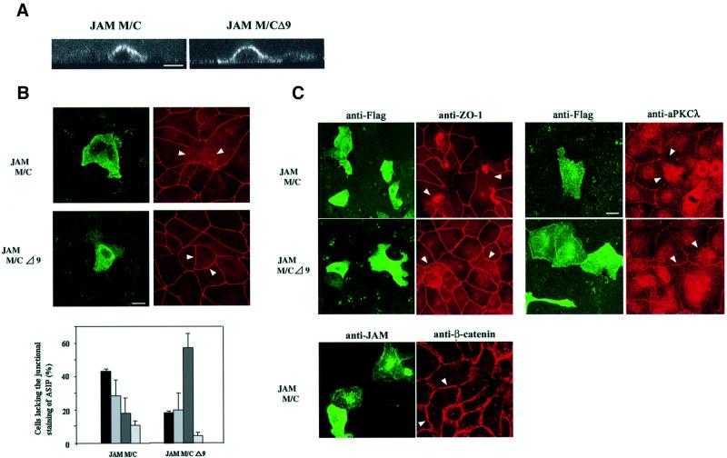
Fig. 7. Overexpression of the cytoplasmic domain of JAM results in the loss of the junctional staining of ASIP in MDCK II cells. Subconfluent MDCK II cells were transiently transfected with expression vectors encoding Flag-tagged mutants of JAM encoding the transmembrane and cytoplasmic domain of JAM, with the complete C-terminus (JAM M/C) or a truncated C-terminus lacking the last nine amino acid residues (JAM M/CΔ9). Cells were immunostained with anti-Flag antibody M2 (A–C) together with anti-ASIP (B), or anti-ZO-1, anti-aPKCλ and anti-β-catenin antibodies (C). (A) Confocal z-sectional views of cells expressing the JAM mutants. Note that both mutants show a similar distribution along the whole cellular surface. (B) JAM-transfected cells appear as bright cells when stained with the anti-FLAG mAb (left panels). Cells overexpressing JAM M/C but not JAM M/CΔ9 lack the junctional staining of ASIP at cell–cell borders (right panels). Based on ASIP localization at cell junctions, cells were classified as complete (complete loss of ASIP, black bars), partial (partial loss, middle grey bars) or intact (intact ASIP staining, dark grey bars). Cells with aberrant shape are represented by light grey bars. Statistical data represent mean values of three independent experiments with a minimum of 200 cells counted for each sample. (C) Cells overexpressing JAM M/C also lack aPKCλ as well as ZO-1 but not β-catenin staining at cell–cell junctions. Note that ZO-1 often forms aggregates in cells expressing JAM M/C but not JAM M/CΔ9. Bars, 15 µm.

An official website of the United States government
Here's how you know
Official websites use .gov
A
.gov website belongs to an official
government organization in the United States.
Secure .gov websites use HTTPS
A lock (
) or https:// means you've safely
connected to the .gov website. Share sensitive
information only on official, secure websites.
