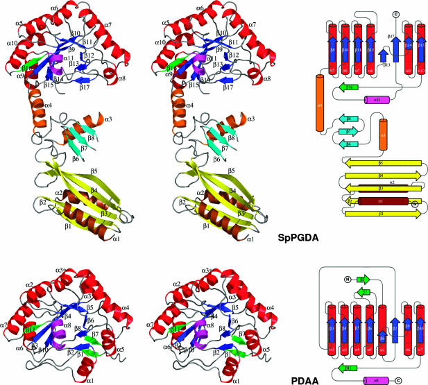
Fig. 1.
SpPgdA structure. Stereo image of the SpPgdA (SpPGDA) and BsPdaA [PDAA, from Protein Data Bank entry 1W1A (11)] structures, alongside topological representations as constructed with topdraw (27). In the SpPgdA N-terminal domain, helices are colored brown and strands, yellow. In the SpPgdA middle domain, helices are colored orange and strands, cyan. In the catalytic domains, helices are colored red and strands, blue, except for the helices (magenta) and strands (green) that do not fit the canonical (β/α)8 fold. Secondary structure elements are named as indicated in the sequence alignment in Fig. 2.

