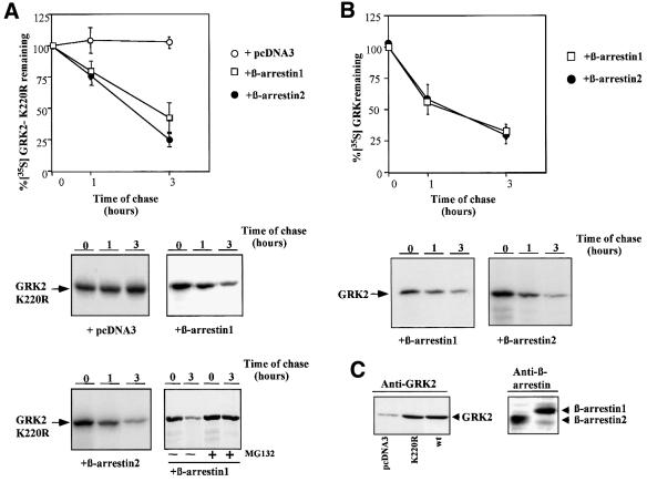
Fig. 2. β-arrestin-1 or β-arrestin-2 overexpression promotes degradation of GRK2-K220R. (A) HEK-293 cells were transiently transfected with GRK2-K220R, β2AR and either β-arrestin-1, β-arrestin-2 or empty vector (pCDNA3), and GRK2-K220R turnover was determined by pulse–chase experiments as detailed in Materials and methods and in Figure 1. Data are the mean ± SE of four or five independent experiments performed in duplicate. A representative gel fluorograph is shown below. The right lower panel shows a similar experiment performed in the presence of the proteasome inhibitor MG132. (B) Similar experiments were performed in cells transiently transfected with wild-type GRK2, β2AR and β-arrestin-1 or β-arrestin-2. Data are the mean ± SE from three or four experiments performed in duplicate, and representative fluorographs are shown below. (C) Lysates from cells transfected with wild-type GRK2 (wt), GRK2-K220R, β-arrestin 1, β-arrestin-2 or empty vector as indicated were subjected to immunoblot analysis with specific GRK2 and β-arrestin antibodies as detailed in Materials and methods.

An official website of the United States government
Here's how you know
Official websites use .gov
A
.gov website belongs to an official
government organization in the United States.
Secure .gov websites use HTTPS
A lock (
) or https:// means you've safely
connected to the .gov website. Share sensitive
information only on official, secure websites.
