
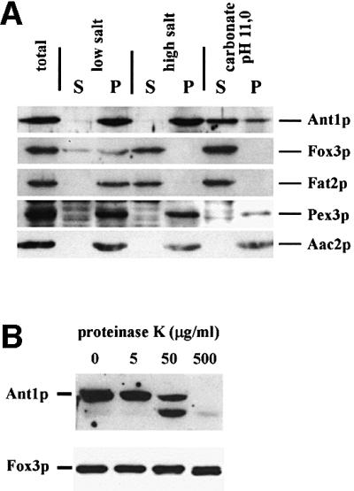
Fig. 3. Ant1p is a peroxisomal membrane protein. (A) Sub-peroxisomal fractionation analysis. A 25 000 g pellet of an oleic acid-induced wild-type strain (UTL-7A) was divided into three parts and treated with either 10 mM Tris–HCl pH 8 (low salt), 10 mM Tris–HCl pH 8/500 mM KCl (high salt) or 100 mM Na2CO3 pH 11 (carbonate). After 30 min incubation, each sample was separated into a pellet (P) and a supernatant (S) fraction by a 200 000 g centrifugation step. Proportionate volumes of the resulting fractions were subjected to immunoblotting using antibodies directed against Ant1p, Pex3p, Fat2p/Pcs60p, Fox3p and Aac2p. (B) Protease protection assay. Organelles isolated from a wild-type strain were split into four parts and incubated for 30 min with increasing concentrations of proteinase K. Reactions were stopped by the addition of 4 mM PMSF and trichloroacetic acid, separated by SDS–PAGE and analyzed by immunoblotting.