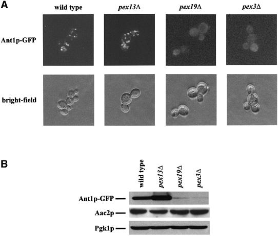
Fig. 4. Peroxisomal location of Ant1p depends on the membrane protein targeting route. (A) Localization of an Ant1p–GFP fusion protein. The wild-type UTL-7A and the otherwise isogenic pex13Δ, pex19Δ and pex3Δ strains expressing Ant1p–GFP under oleic acid-induction conditions were examined for GFP fluorescence. Structural integrity of the cells is documented by bright-field microscopy. (B) Stability of the Ant1p–GFP fusion protein in pex mutants. The same strains as in (A) were induced for 14 h in rich oleate-containing medium. Whole-cell extracts of these samples were analyzed for the amount of Ant1p–GFP, mitochondrial Aac2p and cytosolic Pgk1p by immunological detection.

An official website of the United States government
Here's how you know
Official websites use .gov
A
.gov website belongs to an official
government organization in the United States.
Secure .gov websites use HTTPS
A lock (
) or https:// means you've safely
connected to the .gov website. Share sensitive
information only on official, secure websites.
