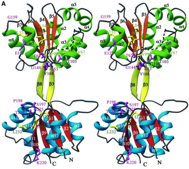
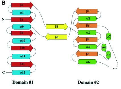
Fig. 2. The U3S structure. Helices and strands of domain 1 are blue and red, helices and strands of domain 2 are green and orange. Strands connecting the two domains are yellow. Secondary structure elements and chain termini are labeled. The dotted line represents the disordered loop (residues 114–118). (A) Ribbon representation of the U3S structure in stereo. The side chains of conserved residues are colored purple if solvent exposed and yellow if buried (see also Figures 3 and 4). (B) Topology diagram. Helices are shown as ellipsoids and strands are shown as arrows. (A) and Figures 4–7 were made using the program Ribbons (Carson, 1991). Secondary structure was defined by DSSP (Kabsch and Sander, 1983).

An official website of the United States government
Here's how you know
Official websites use .gov
A
.gov website belongs to an official
government organization in the United States.
Secure .gov websites use HTTPS
A lock (
) or https:// means you've safely
connected to the .gov website. Share sensitive
information only on official, secure websites.

