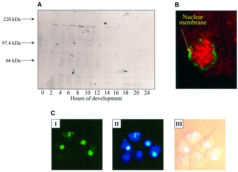
Fig. 3. Localization of PIPkinA protein. (A) Affinity-purified antisera raised against the ankyrin-repeat region of PIPkinA were used to investigate the time course of expression of the PIPkinA protein by western blotting of a developmental time course. Ax2 cells were developed on filters and harvested at the times shown. (B) The staining pattern observed when the affinity-purified antibody against the ankyrin-repeat domain (visualized using rhodamine) was used to stain vegetative amoebae. The green staining indicates the position of the nuclear membrane, revealed using a mouse monoclonal antibody (mab17) that recognizes a component of the nuclear pore apparatus (Fukuzawa and Williams, 1997). (C) A fusion protein containing the PIPkinA coding sequence with GFP fused to the C-terminus was expressed in Dictyostelium cells using a constitutive actin 15 promoter. The localization of the GFP fusion protein in nuclei was observed by fluorescence microscopy (I) and the enrichment in nuclei confirmedby staining with Hoechst 33258 (II). The bright field picture is shown for comparison (III).

An official website of the United States government
Here's how you know
Official websites use .gov
A
.gov website belongs to an official
government organization in the United States.
Secure .gov websites use HTTPS
A lock (
) or https:// means you've safely
connected to the .gov website. Share sensitive
information only on official, secure websites.
