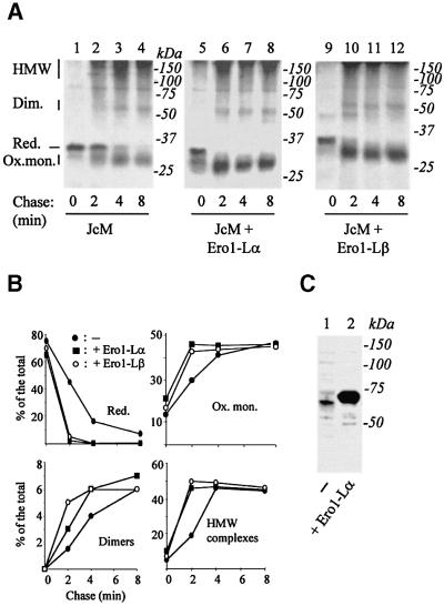
Fig. 1. Ero1-Lα and Ero1-Lβ accelerate oxidative folding of JcM. HeLa cells were co-transfected with pcDNA.3.JcM and pcDNA3.1-ERO1-Lα (lanes 5–8), pcDNA3.1-ERO1-LβHA (lanes 9–12) or an empty vector (lanes 1–4) as a control. Forty-eight hours after transfection, cells were pulsed for 5 min with radioactive amino acids in the presence of DTT (3 mM), washed once at 4°C and chased for the indicated times without the reducing agent. (A) Anti-myc IPs were resolved under non-reducing conditions. The mobility of reduced JcM (Red.), oxidized monomers (Ox. mon.), covalent dimers (Dim.) and high molecular weight complexes (HMW) is indicated on the left-hand margin. (B) The different redox isoforms were quantified by densito metry, and plotted as the per cent of total JcM chains present at each chase time. JcM alone (filled circle); JcM + Ero1-Lα (filled square); JcM +Ero1-Lβ (empty circle). (C) Exogenous Ero1-Lαmyc is expressed at higher levels than endogenous Ero1-Lα. Western blot analysis with anti-Ero1-Lα (D5) from the lysates of 3 × 10⁵ HeLa cells are shown for mock (lane 1) or pcDNA3.1-ERO1-Lαmyc (lane 2) transfected cells. Note the slower mobility of exogenous Ero1-Lα, caused by the presence of a C-terminal tag.

An official website of the United States government
Here's how you know
Official websites use .gov
A
.gov website belongs to an official
government organization in the United States.
Secure .gov websites use HTTPS
A lock (
) or https:// means you've safely
connected to the .gov website. Share sensitive
information only on official, secure websites.
