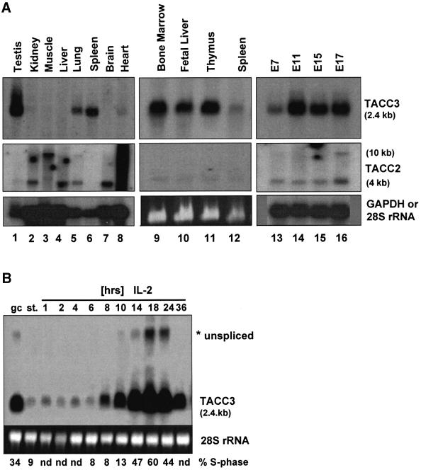
Fig. 1. Lineage-dependent expression of the TACC3 gene. (A) The distribution of TACC3 and TACC2 transcripts in tissues from adult mice (lanes 1–12) and during embryogenesis (days 7–17 of development; lanes 13–16) was determined by northern blot analysis [lanes 1–8 and 13–16, poly(A)+ mRNA; 9–12, total RNA]. (B) Preactivated T cells growing in IL-2 (gc) were starved overnight (st.) and restimulated with IL-2 for the indicated periods of time. TACC3 mRNA levels were analyzed by northern blotting. The percentage of cells in S phase of the cell cycle is indicated (nd, not determined). Hybridization for GAPDH [lanes 1–8 and 13–16 in (A)] or staining of 28S rRNA with ethidium bromide [lanes 9–12 in A, lanes in (B)] was used to control RNA loading.

An official website of the United States government
Here's how you know
Official websites use .gov
A
.gov website belongs to an official
government organization in the United States.
Secure .gov websites use HTTPS
A lock (
) or https:// means you've safely
connected to the .gov website. Share sensitive
information only on official, secure websites.
