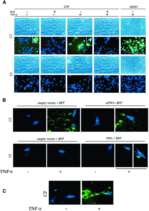
Fig. 5. PW1, but not NFκB, is necessary for TNFα-mediated caspase activation to occur. (A) Proliferating (GM) and differentiating (DM12) cells were subjected to caspase activation analysis (green) and the nuclei stained with Hoechst (blue). Each insert shows an enlarged portion of the corresponding picture. For each microscopic field, the corresponding phase contrast image is also shown. No caspase activity is detected in unstimulated cells (–TNFα), while TNFα (+TNFα) induces caspase activation, both in GM and DM, in C2 cells but not in F3 cells. Successful competition of the non-fluorescent caspase inhibitor (BAF) with the FITC-conjugated caspase substratum demonstrates the specificity of the assay. (B) Caspase activation analysis (green) in cells cotransfected as indicated and identified by the expression of the blue fluorescent protein (BFP, blue). Transfection (empty vector) does not affect caspase activation in C2 cells, both in the absence or presence of TNFα. A dominant-negative form of PW1 (ΔPW1), which inhibits NFκB activation by TNFα, does not affect TNFα-mediated caspase activation. F3 cells are unaffected by transfection procedure alone; however, PW1 expression (PW1) confers the ability to activate caspases when combined with TNFα treatment. (C) C2 cells expressing the NFκB super-repressor IκB show caspase activation upon TNFα treatment, confirming independence of caspase activation from NFκB activation. Data representative of at least two independent experiments are shown.

An official website of the United States government
Here's how you know
Official websites use .gov
A
.gov website belongs to an official
government organization in the United States.
Secure .gov websites use HTTPS
A lock (
) or https:// means you've safely
connected to the .gov website. Share sensitive
information only on official, secure websites.
