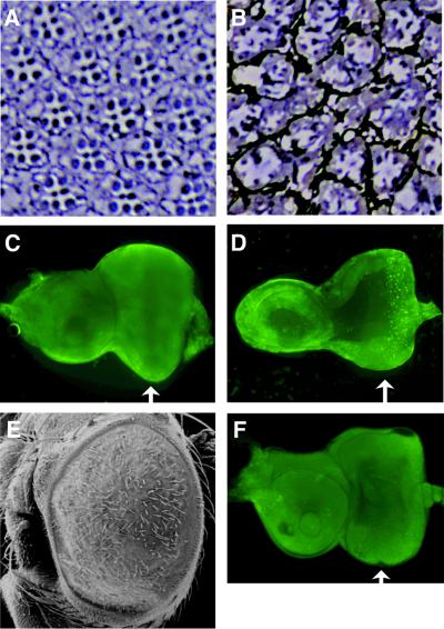
Fig. 5. Decreased number of photoreceptors and induced apoptosis in the GMR–dMybΔC flies. (A and B) Eye sections from wild-type [white; (A)] and GMR–dMybΔC-F25 (B) flies. (C and D) Apoptotic cells were stained with Acridine Orange in wild-type (C) and GMR–dMybΔC-F25 (D) eye discs. Arrows indicate the position of the MF. (E) Scanning electron micrographs of eye of GMR–dMybΔF25 flies expressing p35 ectopically. (F) Apoptotic cells were stained with Acridine Orange in eye discs of GMR–dMybΔF25 flies expressing p35 ectopically.

An official website of the United States government
Here's how you know
Official websites use .gov
A
.gov website belongs to an official
government organization in the United States.
Secure .gov websites use HTTPS
A lock (
) or https:// means you've safely
connected to the .gov website. Share sensitive
information only on official, secure websites.
