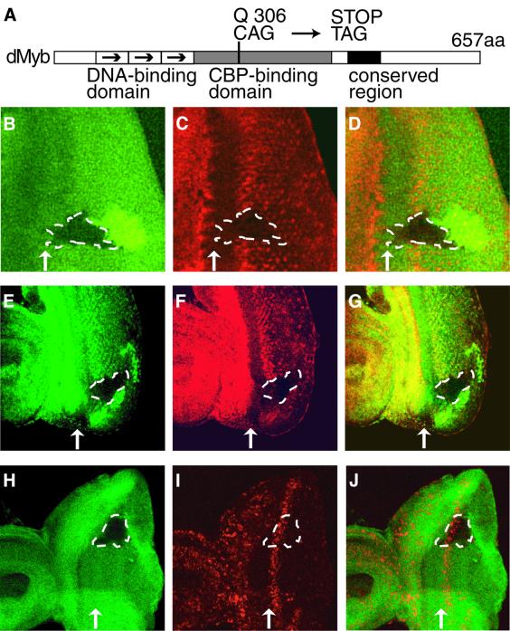
Fig. 6. Loss of cyclin B expression in dmyb-deficient clones. (A) Schematic representation of dMyb mutant. The functional domains and the position of the dmybel1 mutation are indicated. (B–G) Decreased cyclin B expression in dmyb (B–D) or dCBP (E–G) mutant clones in the eye imaginal disc. dmybel1 (B) and nej3 (E) homozygous clones visualized by the lack of β-galactosidase and Myc marker staining (green), respectively, are outlined with dotted lines. Cyclin B expression was monitored in the same disc by staining for the cyclin B protein (red in C and F). The right-hand panels (D and G) show the two single staining patterns superimposed. Anterior is to the left, dorsal is up. At least 10 clones were examined and similar results were obtained with all the clones examined. (H–J) BrdU incorporation of the cells lacking dmyb. dmybel1 (H) homozygous clones visualized by the lack of β-galactosidase marker staining (green) are outlined with dotted lines. BrdU incorporation was monitored in the same disc (red in I and J). The right-hand panel (J) show the two single staining patterns superimposed. Anterior is to the left, dorsal is up. At least 10 clones were examined and similar results were obtained with all the clones examined.

An official website of the United States government
Here's how you know
Official websites use .gov
A
.gov website belongs to an official
government organization in the United States.
Secure .gov websites use HTTPS
A lock (
) or https:// means you've safely
connected to the .gov website. Share sensitive
information only on official, secure websites.
