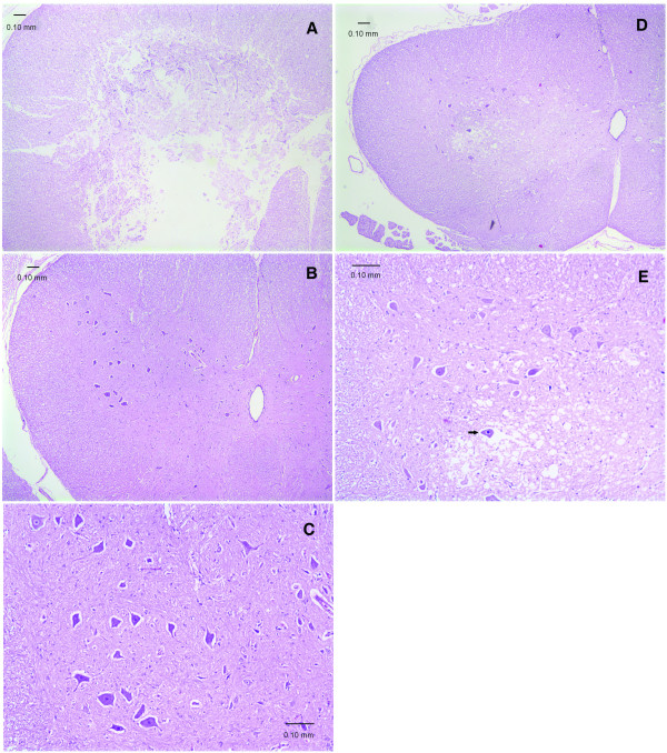
Figure 2.
Transverse sections of rabbit lumbar spinal cord after 1 h ischemia (H&E stain). A. Histopathological score 3, from a rabbit of Group 1. Severe necrosis destroys the entire structure of grey matter indiscriminately. B. Histopathological score 1, from a rabbit of Group 2. No apparent spinal cord damage, the grey matter is well preserved. C. Higher magnification of B showing normal morphology of grey matter and neurons. D. Histopathological score 2, from a rabbit of Group 3. The grey matter is lightly stained with many vacuolations of the neuropil. E. Higher magnification of D. Marked vacuolations of the neuropil in the grey matter, and some neurons are triangular with darkly stained shrunken nuclei (arrow).

