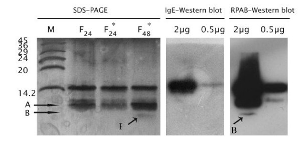
Figure 2.
SDS-PAGE and immunoblot analysis of acid hydrolyzed rCuc m2. silver stained-SDS gel electrophoresis of rCuc m2 after incubation with 75% formic acid for 24 h and 48 h at room temperature (F24) and 37°C (F*24 and F*48). "A" and "B" arrow indicate protein band with molecular mass of approximately 6 and 9 kDa. Figure in the middle shows IgE-immunoblotting of two concentration of F*48 using a pooled serum of melon profilin-sensitized individuals (in the middle) and figure on the right display immunoblotting of the same sample with rabbit polyclonal anti-saffron antibody.

