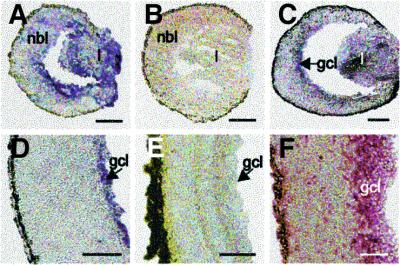
Fig. 1. Representative micrographs of tissue sections of wild-type (Wt1+/+) eyes at different stages of development. Wt1 transcripts are detected by mRNA in situ hybridization in the lens vesicle and retinal neuroblasts of E12 mice (A), as well as in the developing ganglion cell layer of E15 (C) and P1 eyes (D). No Wt1 mRNA is visible in the retinas of adult mice (E) and in retinal tissue of E12 embryos hybridized with digoxigenin-labeled Wt1 sense RNA strand (B). Note, that WT1 protein is detected by immunohistochemistry in the inner portion of the retina obtained at autopsy of a 19-week-gestation human embryo (F). l, lens; nbl, neuroblast layer; gcl, ganglion cell layer. Scale bars, 100 µm.

An official website of the United States government
Here's how you know
Official websites use .gov
A
.gov website belongs to an official
government organization in the United States.
Secure .gov websites use HTTPS
A lock (
) or https:// means you've safely
connected to the .gov website. Share sensitive
information only on official, secure websites.
