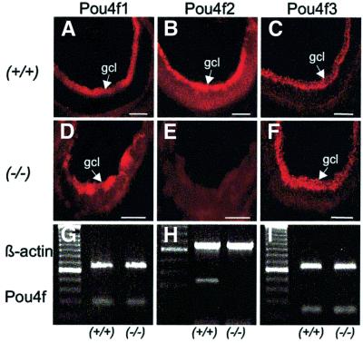
Fig. 4. Expression of Pou4f genes in retinal ganglion cells of wild-type (+/+) and Wt1 null mutant (–/–) retinas at E18. Negative immuno fluorescent staining of the Wt1–/– retina (E) indicates lack of expression of the class IV POU-domain transcription factor Pou4f2. RT–PCR demonstrates absence of Pou4f2 mRNA from the E18 retinas of Wt1–/– embryos (H). No differences are seen in Pou4f1 and Pou4f3 expression (mRNA and protein) between wild-type and Wt1–/– retinas. Shown are representative data obtained from five different embryos each. gcl, ganglion cell layer. Scale bars, 100 µm. A 100 bp DNA ladder was used to estimate the sizes of the expected PCR products. The predicted lengths of the amplified targets are 274 bp (Pou4f1), 300 bp (Pou4f2), 229 bp (Pou4f3) and 642 bp (β-actin), respectively.

An official website of the United States government
Here's how you know
Official websites use .gov
A
.gov website belongs to an official
government organization in the United States.
Secure .gov websites use HTTPS
A lock (
) or https:// means you've safely
connected to the .gov website. Share sensitive
information only on official, secure websites.
