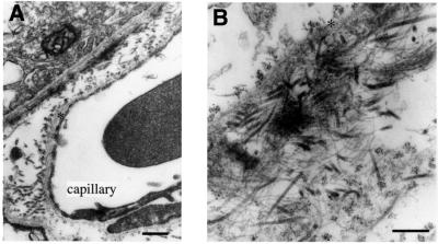
Fig. 9. Immunolabeling of Bruch’s membrane with gold-labeled antibodies against collagen XVIII. (A) Low magnification overview (scale bar 400 nm) shows pigment epithelium in upper left-hand corner, capillary of choriocapillaris at lower right, and clusters of gold particles along lamina densa (asterisks) of epithelial and endothelial basement membranes; label is also seen associated with collagen fibrils in the space between the two basement membranes. (B) Tangential section through Bruch’s membrane shown at a higher magnification (scale bar 250 nm). Clusters of gold particles seen on each side of lamina densa (asterisk) of pigment epithelial cells in upper left-hand corner. At the lower right, clusters of particles seen associated with collagen fibrils. Microfibrils and associated electron-dense material (probably elastin) in central area show little or no labeling.

An official website of the United States government
Here's how you know
Official websites use .gov
A
.gov website belongs to an official
government organization in the United States.
Secure .gov websites use HTTPS
A lock (
) or https:// means you've safely
connected to the .gov website. Share sensitive
information only on official, secure websites.
