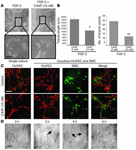
Figure 2.
CA4P impairs capillary tube formation, but has no effect on SMC-stabilized tubes, and destabilizes a preestablished vascular network. (A) CA4P inhibits capillary tube formation. HUVECs (3 × 10⁴) were seeded on Matrigel matrix and incubated in the presence of FGF-2 and with 10 nM CA4P. The effect of CA4P on capillary tube formation was observed after a 12-hour incubation under an inverted light microscope. Magnification: ×10 (top panels), ×40 (bottom panels). Scale bar, 10 μm (top panels), 15 μm (bottom panels). (B) Quantification of the CA4P-mediated capillary tube formation inhibition. The quantification was done by measuring the tubule length and counting the number of branch points in 4 different random pictures. Results are expressed as the mean of the tubule length and the mean of the number of branch points ± SEM (**P < 0.01, #P < 0.001 compared with CA4P-untreated cells; n = 4). (C) SMCs protect capillary tube formation against CA4P. HUVECs (3 × 10⁴) stained with red fluorescent cell linker and SMCs (1 × 10⁴) stained with green fluorescent cell linker were seeded together on Matrigel matrix and incubated in presence of FGF-2 and with 10 nM CA4P. The effect of CA4P on capillary tube formation was observed after a 12-hour incubation under an inverted fluorescent microscope. Magnification: ×20. Scale bar, 50 μm. (D) CA4P destabilizes a preestablished vascular network. HUVECs (3 × 10⁴) were seeded on Matrigel matrix and incubated in presence of FGF-2. Once the capillary network formed (after 12 hours), CA4P was added, and the effect of 10 nM CA4P on the disruption of the capillary network was monitored in a time-course manner every 3 hours. There was significant disruption of endothelial cell morphology (arrows). Magnification: ×40. Scale bar, 10 μm.

