Figure 5.
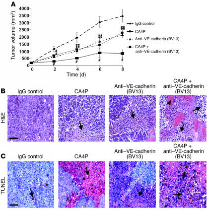
Treatment with CA4P and neutralizing mAb against VE-cadherin (BV13) promotes B16 melanoma tumor necrosis. (A) Growth curves of IgG control–, CA4P-, anti–VE-cadherin– (BV13), or CA4P plus anti–VE-cadherin– (BV13) treated tumors. B16 melanoma cells were injected subcutaneously into the dorsa of C57BL/6 mice. Treatment was initiated after 10 days and mice injected every 2 days. Mice in the CA4P group received an i.v. injection of CA4P at 5 mg/kg; the anti–VE-cadherin group received an i.p. injection of 10 μg of mAb against VE-cadherin (BV13); the combined group received CA4P at 5 mg/kg plus 10 μg of mAb against VE-cadherin (BV13); and the control group received 10 μg of IgG antibody. Tumor size was measured until day 8 after the first injection, and tumor volumes were measured. The data represent the mean value of the tumor volume ± SEM (**P < 0.01, #P < 0.001 compared with IgG control group; n = 9). (B) Histopathological analysis of CA4P- and anti–VE-cadherin– (BV13) treated B16 melanoma tumors. Animals were sacrificed at day 8 after first injection. Shown here are H&E-stained paraffin sections demonstrating areas of necrosis (arrows) with CA4P and mAb against VE-cadherin (BV13) treatment. Arrowhead shows area of hemorrhage. Magnification: ×20. Scale bar, 80 μm. (C) Induction of tumor necrosis. Cell death within paraffin tumor sections was detected by TUNEL. Red staining represents positive signals within the tumors (blue cells are the negative, living cells). Arrows show areas of necrosis. Magnification: ×20. Scale bar, 80 μm.

