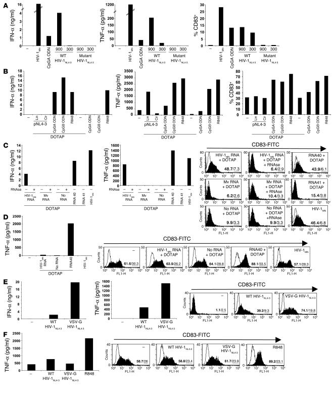
Figure 5.
HIV RNA stimulates pDCs but not MDCs. (A) Flow cytometry–sorted pDCs were exposed overnight to recombinant wild-type or mutant NC SSHS/SSHS HIV-1NL4-3 at the doses of 900 or 300 ng p24CA Eq/ml. As controls, pDCs were cultured with CpGA ODN or HIV-1MN. (B) BDCA-4+ cells were stimulated with 10,000 times more copies of pNL4-3 than the amount present in 900 ng p24CA/ml of HIV-1NL4-3, in the circular (cir) and linearized (lin) forms and after formulation with DOTAP. Low doses of CpGA and CpGB ODNs (1 μg/ml) and R848 (1 μM) were used as controls. Flow cytometry–sorted blood pDCs (C) and MDCs (D) were treated overnight with HIV-1MN RNA, microvesicle-derived RNA (Mv RNA), or with RNA40 and RNA41 mixed with DOTAP as controls. HIV-1MN and Mv RNA were preincubated with RNase prior to formulation with DOTAP. Flow cytometry–sorted pDCs (E) and MDCs (F) were cultured overnight with 900 ng p24CA/ml of wild-type HIV-1NL4-3 or pseudotyped VSV-G HIV-1NL4-3. Concentrations of IFN-α (ng/ml) produced by pDCs and TNF-α (pg/ml) produced by pDCs and MDCs were determined by ELISA. CD83 expression was measured by flow cytometry (the percentage of positive cells is indicated in bold and the mean of fluorescence intensity underlined).

