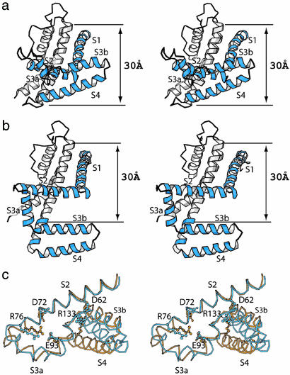
Fig. 2.
Structure of the KvAP-33HI complex and comparison with previous KvAP structures. (a) Stereo view of a single KvAP subunit from the side with extracellular solution above. S1-S4 helices are colored blue. (b) Stereo view of a single KvAP subunit of the KvAP-6E1 Fab complex (PDB ID code 1ORQ). (c) Comparison with the isolated voltage sensor structure. Stereo view of a superposition of the isolated voltage sensor (gold, PDB ID code 1ORS) and the KvAP-Fv complex (blue). The S2 helix (Leu-55 to Tyr-75) was used for superposition, and the S1 helix is not shown. Residues Asp-62, Asp-72, Arg-76, Glu-93, and Arg-133, which are important for channel function, are shown in ball-and-stick representation.

