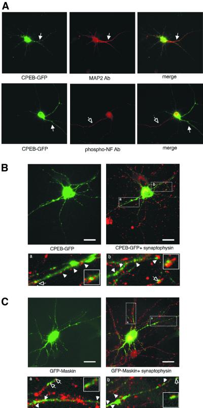
Fig. 3. CPEB–GFP and GFP–maskin in dendrites. (A) CPEB–GFP co-localizes with the dendritic marker MAP2 (solid arrow), but not with the axonal marker phospho-neurofilament (open arrow) in cultured hippocampal neurons. The distribution of CPEB–GFP (B) and GFP–maskin (C) in cultured neurons immunostained with an antibody against the synaptic marker synaptophysin (red, right) is also shown. The magnified images in (A) and (B) show regions of co-localization between CPEB or maskin with synaptophysin (yellow, arrow). The selected synaptic area denoted by the hollow arrow is shown as a high-power image in the inset. Scale bars, 10 µm.

An official website of the United States government
Here's how you know
Official websites use .gov
A
.gov website belongs to an official
government organization in the United States.
Secure .gov websites use HTTPS
A lock (
) or https:// means you've safely
connected to the .gov website. Share sensitive
information only on official, secure websites.
