
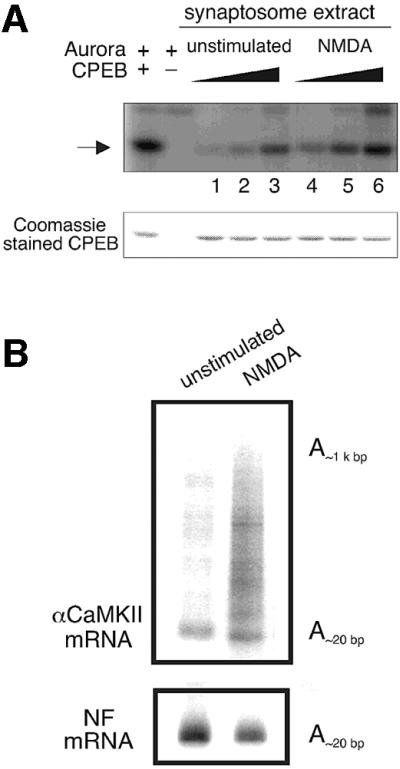
Fig. 7. Activation of NMDA receptor in synaptosomes results in CPEB-phosphorylation and αCaMKII polyadenylation. Unstimulated synaptosomes, or those treated with NMDA as described in Figure 6C, were divided in half; one portion was used for a CPEB phosphorylation experiment (A) while the other portion was used for PAT assays (B). The CPEB phosphorylation (arrow) was measured by using variable amounts of synaptosomal extracts (lanes 1 and 4, 0.1 µl; lanes 2 and 5, 0.5 µl; lanes 3 and 6, 2.5 µl). The Coomassie Blue staining of CPEB shows that equal amounts of substrates were present in all reactions. For the PAT assays, the poly(A) lengths of αCaMKII and neurofilament (NF) mRNA were determined.