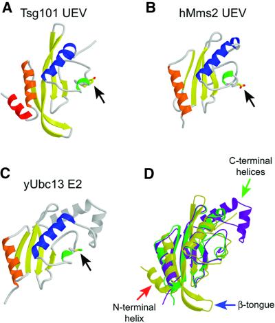
Fig. 2. Structures of E2-fold proteins. (A–C) Ribbon representations of Tsg101 UEV (A), Mms2 (B) and Ubc13 (C). Secondary structure elements are colored as in Figure 1. Black arrows indicate residues that correspond to the active site cysteine of Ubc13. (D) Superposition of the three structures. Tsg101 is colored yellow, Mms2 green and Ubc13 purple. The three major structural differences between Tsg101 and the other two proteins are highlighted by arrows: the extra N-terminal helix (red arrow), the β-hairpin ‘tongue’ (blue arrow) and the missing C-terminal helices (green arrow).

An official website of the United States government
Here's how you know
Official websites use .gov
A
.gov website belongs to an official
government organization in the United States.
Secure .gov websites use HTTPS
A lock (
) or https:// means you've safely
connected to the .gov website. Share sensitive
information only on official, secure websites.
