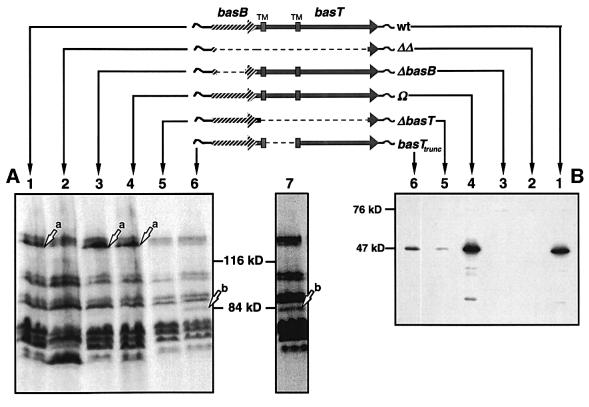
Fig. 3. (A) Patterns of [3H]methyl-labeled proteins of the bas mutant strains in comparison with the wild type. Protein gels were exposed to X-ray films for 1 (lanes 1–6) or 2 (lane 7) weeks. Lanes: 1, wild type; 2, ΔΔ; 3, ΔbasB; 4, Ω; 5, ΔbasT; 6 and 7, basTtrunc. Arrows indicate the positions of BasT (a) and BasTtrunc (b). (B) Immunochemical detection of BasB in total protein extracts using anti-BasB antibodies. Lanes: 1, wild type; 2, ΔΔ; 3, ΔbasB; 4, Ω; 5, ΔbasT; 6 and 7, basTtrunc.

An official website of the United States government
Here's how you know
Official websites use .gov
A
.gov website belongs to an official
government organization in the United States.
Secure .gov websites use HTTPS
A lock (
) or https:// means you've safely
connected to the .gov website. Share sensitive
information only on official, secure websites.
