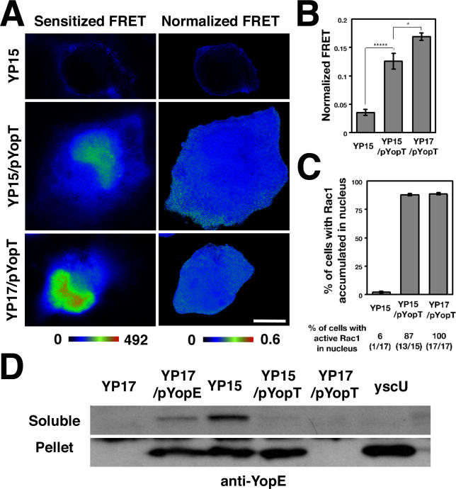
Figure 6. Overexpression of YopT Interferes with YopE-Mediated Inactivation of Rac1 by Blocking YopE Translocation.
COS1 cells were cotransfected with plasmids expressing mYFP-PBD and mCFP-Rac1, challenged for 30 min with Y. pseudotuberculosis strains YP15 (yopE +), YP15/pYopT (yopE +yopT +) or YP17/pYopT (yopE −yopT +) and processed for FRET analysis as in Figure 5 (Materials and Methods).
(A) Typical FRET images of cells incubated with denoted strains.
(B) The presence of pYopT interferes with the ability of YopE to inactivate cytoplasmically localized Rac1. Data represent normalized FRET, using ROIs within the cytoplasms of ten cells. ***** p < 5 × 10⁻⁵; * p = 0.01. Normalized FRET was determined as described (Materials and Methods).
(C) Plasmid-expressed YopT promotes translocation of active Rac1 into the nucleus in the presence of YopE. Nuclear accumulation of Rac1 was determined by presence of mCFP-Rac1 fluorescence (bar graph), and activation was determined in that population showing accumulation using FRET analysis of nuclear regions (data below bar graph; Materials and Methods).
(D) Overexpression of YopT blocks translocation of YopE from bacteria into host cells. After 1 h of infection with Y. pseudotuberculosis strains at MOI = 50, 0.5 × 10⁶ COS1 cells were extracted with 0.1% NP-40 and separated into soluble and pellet fractions to identify translocated YopE in the soluble fraction (Materials and Methods). One-sixth of the soluble fraction and one-half of the pellet were analyzed by immunoblotting using an anti-YopE antibody. Strains used were as in (A), with the addition of the yscU mutant, defective for type III secretion as a negative control.

