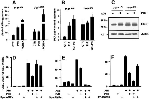
Fig. 5. PrPc-mediated neuroprotective signaling. (A and B) Responses of the cAMP/PKA signaling pathway to the PrR peptide (PrR) in the retina of either wild-type or PrP0/0 mice. Positive controls were either forskolin (FORSK) or a D1-like dopaminergic receptor agonist (6-Chloro-PB). Note the cAMP (A) and PKA (B) responses restricted to wild-type retinal tissue, and the higher basal values in knockout retinas. (C) Western blots for phospho-Elk (top) and loading control with actin (bottom), following treatment of either wild-type or PrP0/0 mouse retinal tissue with the PrR peptide. Note the activation of the Erk pathway restricted to wild-type tissue, as well as the higher basal activity in knockout tissue. (D–F) PrPc-mediated neuroprotective signaling through the cAMP-PKA pathway. Retinal explants from neonatal rats were treated with anisomycin (1 µg/ml), the PrR peptide (PrR 80 µM) in the presence of either 100 µM Rp-cAMP-s (D) or 100 µM Sp-cAMP-s (E), respectively an inhibitor and an activator of cAMP-dependent protein kinase, or in the presence of 30 µM PD98059, an inhibitor of the Erk-activating MEK enzyme (F). Note the reversion of the neuroprotective effect with the PKA inhibitor, and the potentiation of the neuroprotective effect with the Erk pathway inhibitor.

An official website of the United States government
Here's how you know
Official websites use .gov
A
.gov website belongs to an official
government organization in the United States.
Secure .gov websites use HTTPS
A lock (
) or https:// means you've safely
connected to the .gov website. Share sensitive
information only on official, secure websites.
