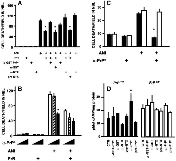
Fig. 6. Effects of antibodies raised against PrPc. (A) Both an antiserum to GST–PrPc (1:80) and an antiserum raised against the neurotoxic peptide (α-NTX) (1:50) prevented neuroprotection by the PrR peptide, while their respective controls, anti-GST and pre-immune serum, had no effect. Data are means ± SEM of triplicate experiments with retinal explants from neonatal rats. *P < 0.01 versus anisomycin alone. (B) A polyclonal antiserum raised against PrPc in PrP0/0 mice blocked anisomycin-induced cell death in the neuroblastic layer, and did not revert the neuroprotective effect of the PrR peptide. In each group of three bars, the concentration of the anti-PrPc antiserum was 0 (open bars), 20 (hatched bars) and 100 (filled bars) µg/ml, respectively. Data are means ± SEM of triplicate experiments with retinal explants from neonatal rats. *P < 0.01 versus no antiserum within the same group. (C) The anti-PrPc antibodies raised in knockout mice (same as in B) had a neuroprotective effect upon retinal explants from wild-type (filled bars), but not from PrP0/0 (open bars) mice. Data are means ± SEM of five replicates in each group. *P < 0.01 versus anisomycin alone among the same genotype. (D) Antibodies that induced neuroprotection increased the intracellular concentration of cAMP in wild-type, but not in PrP0/0 mouse retinal tissue. Results are means ± SEM of three experiments done in triplicate. *P < 0.01 versus control among the same genotype.

An official website of the United States government
Here's how you know
Official websites use .gov
A
.gov website belongs to an official
government organization in the United States.
Secure .gov websites use HTTPS
A lock (
) or https:// means you've safely
connected to the .gov website. Share sensitive
information only on official, secure websites.
