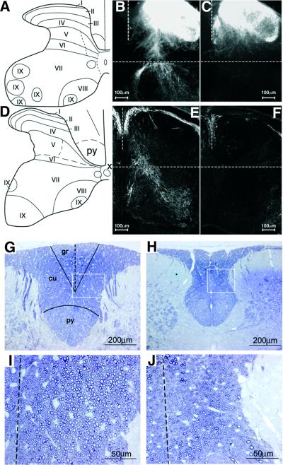
Fig. 4. Analysis of spinal cord proprioceptive afferent projections. (A and D) Schematic diagram of the spinal cord at L4 (A) and C6 (D). I–IX, cytoarchitectonic laminae. (B and C) Anterograde DiI tracing in control (B) and KO (C) mice. Transverse sections of P0 spinal cord at the L4 DRG level. (E and F) Immunostaining of PV in spinal cords of control (E) and KO (F) mice. Transverse sections at C6 DRG level at P0. (G–J) Dorsal column at high cervical level of P30 WT (G) and Runx3 KO (H). Transverse 1 µm epon sections, stained with Toluidine Blue. gr, gracile fascicle; cu, cuneate fascicle; py, pyramidal tract. (I and J) Higher magnification of the squares in (G) and (H), showing a reduced number of large-diameter afferents in the dorsal column of KO mice (J) as compared with WT (I) (the dotted line indicates the midline).

An official website of the United States government
Here's how you know
Official websites use .gov
A
.gov website belongs to an official
government organization in the United States.
Secure .gov websites use HTTPS
A lock (
) or https:// means you've safely
connected to the .gov website. Share sensitive
information only on official, secure websites.
